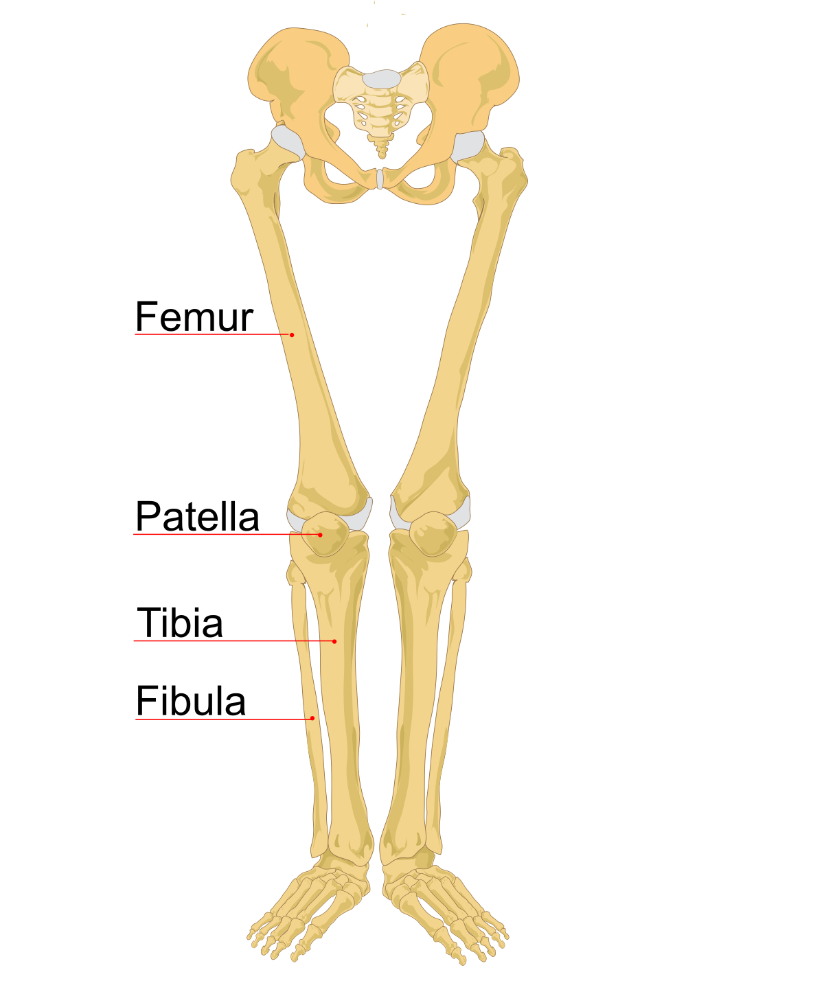
Lower Leg Bone Diagram - leg bones - DriverLayer Search Engine - Start studying leg bone diagram.. The larger bone we refer to as the tibia and is present in front of the lower leg. This can be a difficult fracture to see, because in this case the bones have not moved very far from their correct position. The human leg, in the general word sense, is the entire lower limb of the human body, including the foot, thigh and even the hip or gluteal region. Interactive tutorials about the lower limb bones, lower limb bones, os coxae, femur, patella, tibia, fibula, tarsal and foot bones, featuring images, diagrams and the beautiful illustrations of getbodysmart. Name the 7 bones of the foot (not counting the phalanges).
Anterior view with primary bones names. Health diagram bone skeleton leg knee science anchor chart human human body. Human skeleton long bones of arms and legs britannica. Cheek bone (zygoma) upper jaw (maxilla). The artist's guide to the.
Clipart skeleton skeleton leg, Clipart skeleton skeleton ... from webstockreview.net Knee human anatomy function parts conditions 8 4 bones of the lower limb anatomy and physiology. The two arrows indicate where one of the bones of the leg (the tibia) is broken. Long bone anatomy diagram 12 photos of the long bone anatomy diagram gross anatomy typical long bone diagram, long bone diagram quiz, long bone diagram unlabeled, long bone structure diagram, long. Calcaneus, talus, navicular medial cuneiform, intermediate cuneiform, lateral cuneiform and cuboid. The foot bones shown in this diagram are the talus, navicular, cuneiform, cuboid, metatarsals and calcaneus. Start studying leg bone diagram. Human skeleton long bones of arms and legs britannica. Vtt 150 horse leg anatomy diagram quizlet.
Moreover, the fibula is the smaller bone that goes towards the back part of the leg.
By natalia kremenon january 21, 2021in wiring diagram231 views. The human leg, in the general word sense, is the entire lower limb of the human body, including the foot, thigh and even the hip or gluteal region. Vtt 150 horse leg anatomy diagram quizlet. The knee is a strong but flexible hinge joint. Anterior view with primary bones names. Vector illustration with human skeleton scheme isolated on a white background. Chart of human bones rear view. What is the weight bearing bone of the lower leg? The femur, or thigh bone, is the largest, heaviest, and strongest bone in the human body. At the microscopic level, this hard outer. Bones of the lower limb anatomy and physiology i. The knee joint is the largest joint in the body and is primarily a hinge joint, although some sliding and rotation occur. Posted on april 18, 2019april 18, 2019.
The lower leg has a structure by two bones. The artist's guide to the. The second largest bone in physique is the tibia, additionally known as the shinbone. At the microscopic level, this hard outer. Moreover, the fibula is the smaller bone that goes towards the back part of the leg.
Infographic Diagram Of Human Skeleton Lower Limb Anatomy ... from media.istockphoto.com By natalia kremenon january 21, 2021in wiring diagram231 views. Muscles of the leg and foot classic human anatomy in motion: Related posts of bone anatomy lower leg. Lower bones limbs limb leg diagram muscle foot template anatomy blank human skeleton coloring sketch function th. 2006 kia optima belt diagram. While their parts are similar in general, their structure has been adapted to differing functions. Several muscles attach to and act on the femur. The lower leg has a structure by two bones.
Posted on april 18, 2019april 18, 2019.
Distal end of tibia that forms the medial ankle 15 posterior muscle diagram. Lower jaw (mandible) collar bone. The largest and most medial leg bone, forming both the knee and ankle joints. Your leg bones are the longest and strongest bones in your body. Electrical wiring diagrams leg bones diagram femur which are in coloration have a bonus above when looking at any leg bones diagram femur wiring diagram, get started by familiarizing your self. Vector illustration with human skeleton scheme isolated on a white background. By natalia kremenon january 21, 2021in wiring diagram231 views. Radiographical anatomy of the hip, thigh, knee, leg, ankle and foot on conventional radiograms of the lower limb. Vtt 150 horse leg anatomy diagram quizlet. Posted on january 21, 2015 by admin. Long bone anatomy diagram 12 photos of the long bone anatomy diagram gross anatomy typical long bone diagram, long bone diagram quiz, long bone diagram unlabeled, long bone structure diagram, long. Fractures of the bones of the lower leg (the tibia and fibula). Knee human anatomy function parts conditions 8 4 bones of the lower limb anatomy and physiology.
Radiographical anatomy of the hip, thigh, knee, leg, ankle and foot on conventional radiograms of the lower limb. By natalia kremenon january 21, 2021in wiring diagram231 views. Vector illustration with human skeleton scheme isolated on a white background. Ankle human anatomy image function conditions more. Labeled diagram of long bone.
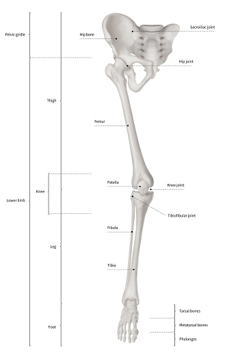
Bones of the Lower Limb Unlabeled | Anatomy bones, Anatomy ... from i.pinimg.com Ankle and foot bones and joints unit 4/12/18 lower leg: The knee joint is the largest joint in the body and is primarily a hinge joint, although some sliding and rotation occur. At the microscopic level, this hard outer. Dog leg bones diagram wiring schematic diagram www. The bones involved in it, however, are only the femur and the tibia, although the. Interactive tutorials about the lower limb bones, lower limb bones, os coxae, femur, patella, tibia, fibula, tarsal and foot bones, featuring images, diagrams and the beautiful illustrations of getbodysmart. The knee is a strong but flexible hinge joint. It is the tibial joint surface or ceiling of the ankle mortise.
Bones of the leg and foot, lower leg bone anatomy, leg bones anatomy, leg muscles, leg bones diagram, leg bone structure, leg anatomy muscles, parts of the lower leg.
Standard radiography view of anatomical structures of the lower limb. It is the tibial joint surface or ceiling of the ankle mortise. Lower jaw (mandible) collar bone. At the microscopic level, this hard outer. The lower leg has a structure by two bones. The knee joint is the largest joint in the body and is primarily a hinge joint, although some sliding and rotation occur. Dog leg bones diagram wiring schematic diagram www. Bones of the lower limb anatomy and physiology i. Labeled diagram of long bone. The largest and most medial leg bone, forming both the knee and ankle joints. Several muscles attach to and act on the femur. Normal leg bones are relatively straight, but those affected by paget's disease are porous and figure 9. By natalia kremenon january 21, 2021in wiring diagram231 views.
The two arrows indicate where one of the bones of the leg (the tibia) is broken leg bone diagram. Lower bones limbs limb leg diagram muscle foot template anatomy blank human skeleton coloring sketch function th.


0 Komentar